High-Frequency Ultrasound: The Most Important Diagnostic Tool in Thyroid Care
Written by John P. Sabra, MD FACS
Updated May 2026
Educational only. This article is not medical advice. Always consult your physician about your individual situation.
No imaging technology has done more to transform thyroid diagnosis and treatment than ultrasound. Understanding why — and what separates excellent thyroid ultrasound from ordinary neck imaging — helps patients ask the right questions at every stage of their care.
What High-Frequency Ultrasound Is
Standard ultrasound used in general medical imaging typically operates at 2 to 5 megahertz. High-frequency ultrasound for thyroid and neck evaluation operates at 10 to 18 megahertz or higher. Higher frequency sound waves produce shorter wavelengths and generate finer detail — at the cost of depth penetration. The thyroid sits just beneath the skin of the lower neck, rarely more than 2 to 3 centimeters deep, making it ideal for high-frequency imaging.
The practical result: an image quality that allows the operator to see structures as small as 2 to 3 millimeters with clarity, to distinguish solid from cystic tissue, to evaluate blood flow using Doppler, to identify calcium deposits smaller than a millimeter, and to assess nodule margins with enough precision to characterize them as smooth, irregular, or spiculated — the features that drive the TIRADS score and the clinical decision making that follows.
Why Ultrasound Is the Gold Standard for Thyroid Evaluation
CT scanning provides excellent anatomical detail and is indispensable for evaluating substernal goiters and large cancers, but uses ionizing radiation and cannot characterize a thyroid nodule’s internal features with the resolution that ultrasound provides. MRI avoids radiation but is slower, more expensive, and generally inferior to high-quality ultrasound for nodule characterization. Neither modality can guide a biopsy needle in real time with the immediacy and precision of ultrasound.
High-Frequency Ultrasound in the Clinic
In a dedicated thyroid practice, high-frequency ultrasound is not a test ordered and sent to a radiology department. It is performed by the physician in the room, interpreted in real time, and integrated directly into the clinical decision. When a thyroid specialist performs ultrasound during your clinic visit, several things happen that do not happen when you are scanned by a technologist and read by a radiologist who never meets you:
- The physician correlates what they see on the screen with what they feel on physical examination
- They direct the probe toward a specific area of concern in real time
- They compare the current scan immediately against prior imaging in their system
- They can explain what they are seeing to the patient as they scan
- They can proceed directly to a biopsy or other intervention if findings warrant it — without scheduling a separate appointment
High-Frequency Ultrasound in the Operating Room
The role of ultrasound does not end when the patient moves from clinic to operating room. Intraoperative ultrasound has become a valuable tool in thyroid and parathyroid surgery for several specific applications:
- Locating small nodules during surgery — a nodule clearly visible on a preoperative scan may be difficult to identify by palpation alone once the neck is open
- Identifying suspicious lymph nodes intraoperatively — nodes not visible on preoperative imaging may become apparent when the surgeon can scan directly with the probe at the operative field
- Parathyroid localization during parathyroid surgery — confirming the location of an adenoma before the incision and during dissection, reducing operating time
- Confirming adequate resection — a quick intraoperative scan confirms the area of concern has been addressed before the wound is closed
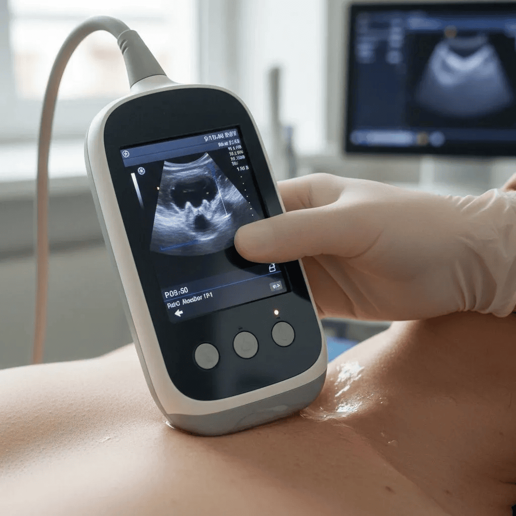
Modern high-frequency ultrasound technology has become remarkably compact. Portable units the size of a tablet, and handheld probes that connect to a smartphone, are now capable of producing diagnostic-quality thyroid images. This portability has extended the reach of thyroid ultrasound into clinic rooms, bedside consultations, and outpatient procedure suites where dedicated imaging equipment was not previously available.
What to Look for in a Thyroid Ultrasound Evaluation
The standard worth looking for is a physician who performs their own ultrasound at the point of care using a high-frequency dedicated thyroid probe, who interprets the images in the context of the full clinical picture, and who can move directly from imaging to a biopsy or treatment plan without an intervening radiology step.
Ask your physician: do you perform the ultrasound yourself during my appointment, or is it done separately in radiology? The answer tells you a great deal about how integrated the imaging is with the clinical care, and how directly the physician’s own eyes are involved in what they recommend for your nodule.
References
- Tessler FN, et al. ACR TI-RADS White Paper. J Am Coll Radiol. 2017.
- Haugen BR, et al. 2015 ATA Management Guidelines for Thyroid Nodules and DTC. Thyroid. 2016.
- ACR TI-RADS — Thyroid Imaging Reporting and Data System.
This article was written by John P. Sabra, MD FACS and is intended for patient education only. It does not constitute medical advice and does not replace a consultation with your physician.
More from the Technology and Tools series


Ready to Talk to a Specialist?
A thyroid specialist can review your specific results, answer your questions, and help you understand exactly what your next steps should be. The ATLAS network connects you with experienced thyroid and parathyroid expert physicians who focus on thyroid disease.
Find a Thyroid Specialist Near You →