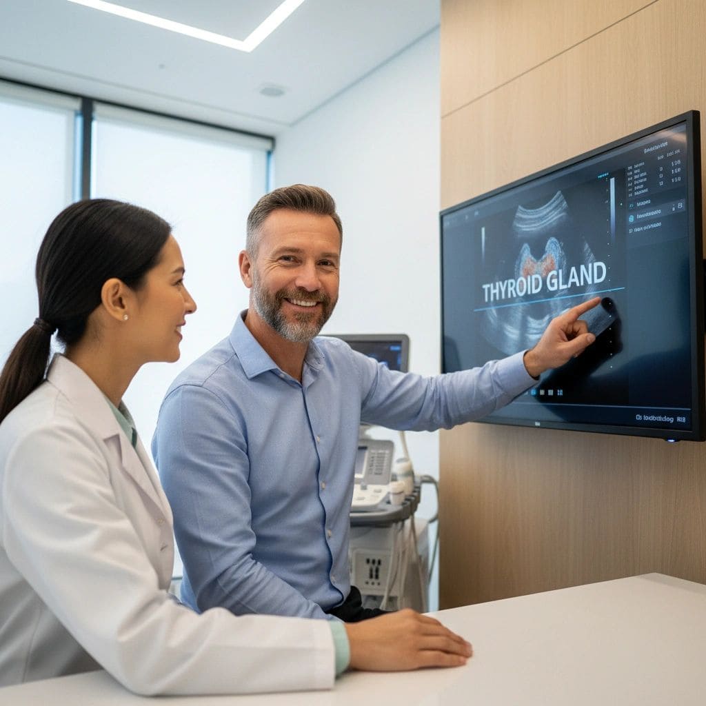
Highly Suspicious Thyroid Nodule (TIRADS 5 / TR5): What That Means and What Comes Next
Written by John P. Sabra, MD FACS
Updated May 2026
Educational only. This article is not medical advice. Always consult your physician about your individual situation.
If your ultrasound report says TIRADS 5, or TR5, and you have seen the words “highly suspicious,” take a moment before you go further down the internet rabbit hole. This article is written specifically for where you are right now.
Here is the most important fact on this page: a TR5 result is not a cancer diagnosis. It means your radiologist identified several ultrasound features that are associated with a higher likelihood of malignancy. It means a biopsy is the essential and urgent next step. What it does not mean is that you have cancer, that surgery is inevitable, or that the outcome is determined. The majority of TR5 nodules that proceed to biopsy are still benign.
The goal of this article is not to minimize the seriousness of a TR5 result — it warrants prompt attention and a specialist consultation. The goal is to give you an accurate, calm picture of what this category means, what happens next, and what the realistic range of outcomes looks like.
TIRADS Is a Framework, Not a Diagnosis
TIRADS is a scoring framework developed by the American College of Radiology to standardize how radiologists communicate risk. The scale runs from TR1 (normal) to TR5 (highly suspicious). It is built on population-level evidence and is one of the most useful tools available for guiding thyroid nodule management.
But it is a framework, not a diagnosis. A TIRADS score tells your physician what the ultrasound features suggest about probability — it does not tell them what is definitively happening inside your nodule. Only a tissue biopsy can do that.
Where TR5 Sits and What “Highly Suspicious” Actually Means
| Level | Description | Malignancy Risk | ACR Guideline FNA Threshold |
|---|---|---|---|
| TR1 | Benign | 0% | Not typically indicated |
| TR2 | Not suspicious | <1% | Not typically indicated |
| TR3 | Mildly suspicious | ~5% | Consider FNA at ≥ 2.5 cm |
| TR4 | Moderately suspicious | 6–17% | Consider FNA at ≥ 1.5 cm |
| TR5 ▶ | Highly suspicious | 20–35%+ | Consider FNA at ≥ 1.0 cm — patient factors may shift this |
TR5 carries an estimated malignancy risk of roughly 20 to 35% in published clinical studies. Read that again carefully: in the range of 65 to 80% of TR5 nodules, a biopsy comes back benign. “Highly suspicious” means the features are concerning enough to warrant prompt evaluation, not that cancer has been found.
The reason biopsy is so important at TR5 is precisely because the stakes of not knowing are higher. A 20 to 35% malignancy probability is meaningful, and the consequence of missing a cancer that could have been caught early is far greater than the minor discomfort of an office biopsy. This is the category where acting promptly genuinely matters.
The Ultrasound Features That Create a TR5 Score
TR5 is not a single finding — it is a constellation of features that together push a nodule into the highest-suspicion tier.
| Feature | What TR5 Looks Like | Plain-English Meaning |
|---|---|---|
| Echogenicity | Markedly hypoechoic | The nodule appears significantly darker than the surrounding muscle tissue on ultrasound — a strong association with malignancy. |
| Shape | Taller than wide | The nodule is taller than it is wide when measured in cross-section. This orientation is more common in cancerous growth. |
| Margins | Irregular or spiculated | The edges of the nodule are jagged, angular, or have spike-like projections rather than smooth, clean borders. |
| Echogenic foci | Microcalcifications | Tiny, bright flecks of calcium within the nodule. These punctate dots are one of the more specific ultrasound markers for papillary thyroid cancer. |
| Extrathyroidal extension | Invasion beyond thyroid capsule | The nodule appears to extend beyond the outer edge of the thyroid gland. This is the highest-concern finding and is relatively uncommon. |
What the FNA Recommendation Looks Like for TR5
Per ACR guidelines, FNA biopsy is recommended for TR5 nodules that are 1.0 cm or larger.
- Nodule at or above 1.0 cm: Biopsy is recommended promptly. The clinical consensus at TR5 for acting at 1.0 cm is strong.
- Nodule between 0.5 and 1.0 cm: Follow-up imaging at 6 to 12 months is the typical starting point. If the nodule grows or develops new features, biopsy is then recommended.
- Nodule under 0.5 cm: Generally not biopsied regardless of features, given the extremely low likelihood of a clinically meaningful cancer at that size.
FNA is a brief, well-tolerated office procedure. A thin needle is guided into the nodule under real-time ultrasound imaging, cells are collected for pathology review, and the whole process takes approximately 20 to 30 minutes. No general anesthesia, no hospital admission, no incision. Results return within three to seven business days, categorized using the standardized Bethesda classification system.
If Your Biopsy Comes Back Positive: What Happens Next
If your FNA result confirms malignancy, the most important thing to understand immediately is this: thyroid cancer is one of the most treatable cancers that exists.
| Step | What Happens | What to Know |
|---|---|---|
| FNA Biopsy | Cells sampled under ultrasound guidance | A brief office procedure under local anesthetic. Results in 3 to 7 business days. |
| Surgical Referral | Referral to a thyroid surgeon for evaluation | The surgeon reviews your imaging, biopsy result, and overall health to determine the appropriate surgical approach. |
| Surgery Options | Lobectomy (half thyroid) or total thyroidectomy | Many low-risk thyroid cancers are now managed with lobectomy alone, preserving partial thyroid function and often avoiding lifelong hormone therapy. |
| Survival Context | Prognosis for most patients is excellent | Papillary thyroid cancer — the most common type found in TR5 nodules — has a five-year survival rate exceeding 98% for most patients. |
Not all thyroid cancer requires total thyroidectomy. Many patients with low-risk papillary thyroid cancer are now managed with lobectomy: removal of only the affected half of the thyroid. This often preserves enough thyroid function to avoid lifelong hormone replacement — a significant improvement in quality of life compared to older treatment approaches.
A Note on RFA: Why It Is Not the Right Path for TR5
If you have been researching thyroid treatment options, you may have come across radiofrequency ablation (RFA) — a non-surgical procedure that uses heat to shrink thyroid nodules. It is a legitimate and effective treatment for confirmed benign nodules.
RFA is not appropriate for TR5 nodules. RFA treats the nodule from the inside; it does not produce a tissue sample, does not confirm whether cancer is present, and does not address the lymph nodes or surrounding structures that surgical evaluation would include. Using RFA on a high-suspicion nodule without a confirmed benign biopsy would mean treating an unknown — potentially allowing a cancer to persist without definitive treatment.
The sequence for TR5 is: biopsy first. If the result is benign, the full range of treatment options (including surveillance, RFA, or surgery) can then be discussed. If the result is malignant, a surgical plan is built from there.
The Bottom Line for TR5 Patients
A TR5 result is the highest-concern TIRADS category, and it warrants prompt, serious attention. Schedule a biopsy soon and arrange a specialist consultation either before or immediately after. That is not an overreaction — it is the appropriate, evidence-based response to a high-suspicion finding.
What it is not is a diagnosis. The majority of TR5 nodules that are biopsied still come back benign. And for the minority that are confirmed malignant, the treatment pathway for thyroid cancer is well-established, the outcomes are among the best of any cancer type, and the options available today — including nerve-sparing surgery and thyroid-preserving lobectomy — are far better than they were even a decade ago.
The most important thing you can do right now is get to the right specialist: a high-volume thyroid endocrinologist or thyroid surgeon who reviews cases like yours regularly, with your full ultrasound report in hand and a clear understanding of your specific feature profile. Act promptly.
References
- Tessler FN, et al. ACR TI-RADS White Paper. J Am Coll Radiol. 2017.
- Haugen BR, et al. 2015 ATA Management Guidelines for Thyroid Nodules and DTC. Thyroid. 2016.
- National Cancer Institute — Thyroid Cancer.
This article was written by John P. Sabra, MD FACS and is intended for patient education only. It does not constitute medical advice and does not replace a consultation with your physician. TIRADS categories are clinical guidelines, not mandatory rules. Individual nodule characteristics, patient health history, age, risk factors, and clinical judgment all determine the appropriate management for every patient.
More from the Ultrasound series


Ready to Talk to a Specialist?
A thyroid specialist can review your specific results, answer your questions, and help you understand exactly what your next steps should be. The ATLAS network connects you with experienced thyroid and parathyroid expert physicians who focus on thyroid disease.
Find a Thyroid Specialist Near You →