How to Read Your Thyroid Ultrasound Report: A Plain-English Guide
Written by John P. Sabra, MD FACS
Updated May 2026
Educational only. This article is not medical advice. Always consult your physician about your individual situation.
Most patients receive their thyroid ultrasound report the same way: through a patient portal notification, often late in the evening, often before their doctor has had a chance to review it with them. The report sits on the screen full of unfamiliar terms. Hypoechoic. Spiculated. TIRADS 4. Echogenic foci. It is written for radiologists, not patients, and it can turn a routine imaging result into an anxious night of searching.
This article is a practical guide to decoding that report. It covers every major section you will encounter on a standard thyroid ultrasound report, explaining what each term means, why it matters, and how it connects to the overall picture of your thyroid health. You do not need a medical background to understand your results. You need someone to translate them, and that is exactly what this article does.
Bookmark this page. Come back to it before your next appointment. Use it alongside the Atlas Ultrasound Analysis tool to build a clear, specific picture of your individual result, and arrive at your specialist appointment ready to ask the right questions.
What a Thyroid Ultrasound Actually Measures
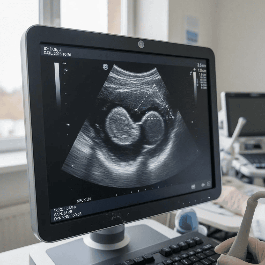
A thyroid ultrasound uses high-frequency sound waves to create a detailed image of your thyroid gland and any nodules within it. Unlike an X-ray, it involves no radiation. Unlike an MRI, it is inexpensive, widely available, and can be done in a physician’s office. It is the single most important first-line tool for evaluating thyroid nodules.
A standard thyroid ultrasound report will document the following for each nodule identified:
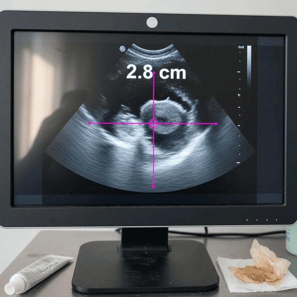
- Size: Measured in three dimensions (length, width, and depth), typically in centimeters. Size matters for determining whether biopsy is recommended, though it does not directly predict malignancy risk on its own.
- Location: Which lobe of the thyroid the nodule sits in (right, left, or isthmus), and its position within that lobe.
- Composition: Whether the nodule is solid, cystic (fluid-filled), mixed, or spongiform.
- Echogenicity: How the nodule’s brightness compares to surrounding thyroid tissue, ranging from markedly hypoechoic to hyperechoic.
- Shape: Whether the nodule is wider than it is tall, or taller than it is wide, as measured in the cross-sectional view.
- Margins: The character of the nodule’s edges: smooth, ill-defined, irregular, or spiculated.
- Echogenic foci: Any bright spots within the nodule, including microcalcifications, comet-tail artifacts, or rim calcifications.
- TIRADS / TR score: The overall suspicion category assigned based on the cumulative point total from the above features.
Each of these elements contributes to the complete picture. No single feature tells the whole story; it is the combination that drives the score and the management recommendation.
The Five Features That Drive Your TIRADS Score
The ACR TIRADS scoring system assigns points to five specific ultrasound features. Those points are added together to produce a final TR score from 1 to 5.
| Feature | What You’ll See | What It Means |
|---|---|---|
| Composition | Solid, cystic, spongiform, or mixed | Solid nodules contain more cells. Fully fluid-filled (cystic) nodules are almost always benign. Spongiform nodules are considered essentially benign. |
| Echogenicity | Hypoechoic, isoechoic, hyperechoic, or markedly hypoechoic | Isoechoic and hyperechoic appearances are reassuring. Hypoechoic and markedly hypoechoic carry progressively higher concern. |
| Shape | Wider than tall, or taller than wide | A nodule that is taller than it is wide is one of the stronger individual markers for malignancy. |
| Margins | Smooth, ill-defined, irregular, or spiculated | Smooth and ill-defined margins are low risk. Irregular or spiculated margins are higher-risk features. |
| Echogenic foci | Microcalcifications, comet-tail artifacts, or rim calcifications | Microcalcifications are one of the most specific markers for papillary thyroid cancer. Comet-tail artifacts are benign. Rim calcifications carry intermediate concern. |
The TIRADS Scoring System: What Each Level Means
Once the five features have been scored and added together, the total determines your TR level from TR1 to TR5.
| Level | Description | Malignancy Risk | FNA Threshold | Typical Next Step |
|---|---|---|---|---|
| TR1 | Benign | 0% | None | No follow-up needed |
| TR2 | Not suspicious | <1% | None | No follow-up typically needed |
| TR3 | Mildly suspicious | ~5% | ≥ 2.5 cm | Surveillance ultrasound at 1, 3, and 5 years |
| TR4 | Moderately suspicious | 6–17% | ≥ 1.5 cm | Surveillance or FNA depending on size and clinical context |
| TR5 | Highly suspicious | 20–35%+ | ≥ 1.0 cm | Prompt FNA biopsy and specialist consultation |
Two points are important to understand about this table. First, the FNA thresholds shown are guidelines, not rules. A physician may recommend biopsy below the size threshold for clinical reasons, including symptoms, patient preference, growth on surveillance, or candidacy for a non-surgical treatment such as RFA. The table describes what guidelines suggest for the average patient. Your physician applies those guidelines to you as an individual.
Second, the malignancy risk figures shown are population-level estimates from published studies. A TR4 nodule with 4 points at the lower end of the range carries a very different practical risk than one with 6 points at the upper end, even though both appear in the same row of this table.
Common Questions Patients Bring to Their First Appointment
After reviewing reports with patients over many years, a handful of questions come up again and again. Here are the most common ones, answered directly.
“My nodule is 2 cm. Is that large?”
In the context of thyroid nodules, 2 cm is moderate in size, not small and not particularly large. What matters far more than the raw measurement is the TIRADS score produced by the nodule’s features. A 2 cm nodule that is isoechoic, smooth-margined, and has no microcalcifications carries a very different risk profile than a 1 cm nodule that is markedly hypoechoic with irregular margins. Size matters primarily because it determines the FNA threshold for your specific TIRADS category, not because it independently predicts malignancy.
“My report says I have multiple nodules. Is that worse?”
Not necessarily. A multinodular thyroid is extremely common and does not in itself increase overall malignancy risk beyond what each individual nodule’s features would suggest. Per ACR guidelines, biopsy is generally limited to the two nodules with the most suspicious features, not every nodule present. The most suspicious nodule, not the largest, is what drives management decisions.
“My report says the nodule is hypoechoic. Should I be worried?”
Hypoechoic simply means the nodule appears darker than the surrounding thyroid tissue on ultrasound. It is one factor among five in the TIRADS scoring system. On its own, being hypoechoic adds 2 points to the score. It is a feature worth noting, but not a cause for immediate alarm. A hypoechoic nodule with smooth margins, a wider-than-tall shape, and no microcalcifications is a very different finding than a hypoechoic nodule with irregular margins and microcalcifications.
“My report uses different terminology than what I have read online.”
This is common and understandably confusing. The ACR TIRADS system described in this article is the most widely used framework in the United States, but other systems exist. Some reports use American Thyroid Association risk categories rather than TIRADS numbers. If your report does not include a TR score or a clear risk category, ask your physician which system was used.
Quick-Reference Glossary: Terms You Will See on Your Report
The following table covers the terms that appear most frequently on standard thyroid ultrasound reports. Use it as a reference alongside your own report.
| Term | Plain-English Meaning |
|---|---|
| Nodule | A discrete lump or growth within the thyroid gland that is radiologically distinct from the surrounding tissue on ultrasound. |
| Echogenicity | How bright or dark the nodule appears compared to the normal thyroid tissue surrounding it. |
| Hypoechoic | Darker than the surrounding thyroid tissue. The darker the nodule appears, the higher the level of concern. |
| Isoechoic | The same brightness as the surrounding thyroid tissue. Generally reassuring. |
| Hyperechoic | Brighter than the surrounding thyroid tissue. Also generally reassuring. |
| Spongiform | Composed of many tiny fluid-filled spaces. Almost always benign. |
| Microcalcifications | Tiny, punctate bright flecks within the nodule. One of the more specific ultrasound markers for papillary thyroid cancer. |
| Taller than wide | A nodule whose height exceeds its width when viewed in cross-section. A higher-risk feature. |
| Extrathyroidal extension | The nodule appearing to extend beyond the outer capsule of the thyroid gland. A high-concern finding. |
| FNA | Fine Needle Aspiration. A needle biopsy performed under ultrasound guidance to collect cells for pathology review. |
| TIRADS / TR score | Thyroid Imaging Reporting and Data System. A 1–5 scale summarizing overall suspicion level based on combined ultrasound features. |
| ACR | American College of Radiology. The organization that developed and maintains the TIRADS scoring system. |
The Bottom Line
A thyroid ultrasound report is not a verdict. It is a description. Every term on the page is a radiologist’s objective observation of what your nodule looks like under imaging. Those observations are translated into a risk score, and that score is a starting point for a clinical conversation, not a definitive diagnosis.
The most useful thing you can do with your report right now is understand it feature by feature, not just react to the final score. Know what your composition finding means. Know whether your margins are smooth or irregular. Know whether microcalcifications were identified. That level of understanding transforms your next physician appointment from a passive experience into an active one.
Use the Atlas tool to walk through your specific features and see how your score is built. Then bring that analysis to a specialist who can apply clinical judgment to your complete picture and help you build a care plan that is designed around you, not a population-level guideline.
References
- Tessler FN, et al. ACR Thyroid Imaging, Reporting and Data System (TI-RADS): White Paper. J Am Coll Radiol. 2017.
- Haugen BR, et al. 2015 ATA Management Guidelines for Adult Patients with Thyroid Nodules and Differentiated Thyroid Cancer. Thyroid. 2016.
- American Thyroid Association — Thyroid Nodules: Patient Information.
This article was written by John P. Sabra, MD and is intended for patient education only. It does not constitute medical advice and does not replace a consultation with your physician. TIRADS categories are clinical guidelines, not mandatory rules. Individual nodule characteristics, patient health history, age, risk factors, and clinical judgment all determine the appropriate management for every patient.
More from the Ultrasound series


Ready to Talk to a Specialist?
A thyroid specialist can review your specific results, answer your questions, and help you understand exactly what your next steps should be. The ATLAS network connects you with experienced thyroid and parathyroid expert physicians who focus on thyroid disease.
Find a Thyroid Specialist Near You →