Parathyroid Adenoma: The Most Common Cause of Hyperparathyroidism
Written by John P. Sabra, MD FACS
Updated May 2026
Educational only. This article is not medical advice. Always consult your physician about your individual situation.
In roughly 85% of patients with primary hyperparathyroidism, the cause is a single small benign tumor growing on one of the four parathyroid glands. It is called a parathyroid adenoma. It is not cancer, it is not dangerous on its own, but it is the reason the calcium is high, the bones are thinning, and the patient feels unwell.
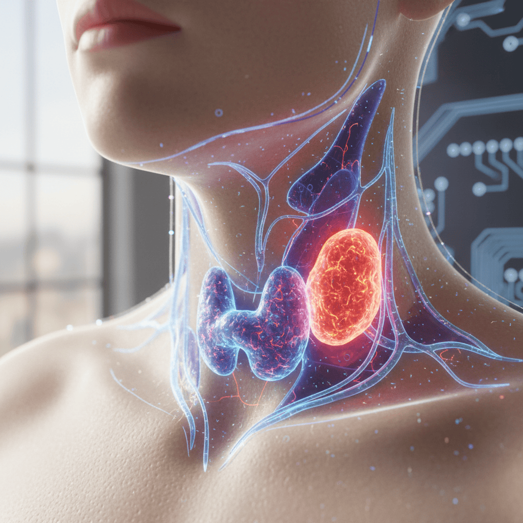
A parathyroid adenoma is a benign growth on one of the four parathyroid glands in the neck. The term adenoma simply means a benign tumor of glandular tissue. A parathyroid adenoma is composed mostly of the same type of cells that make up a normal parathyroid gland, called chief cells, but they have escaped the normal feedback control that tells them when to stop producing parathyroid hormone.
A normal parathyroid gland produces PTH only when calcium is low. When calcium rises, the gland senses it through a receptor on its surface (the calcium-sensing receptor) and turns PTH production down. An adenoma has a defect in that feedback system. It continues to produce PTH regardless of the calcium level, as though the thermostat has become stuck. The result is chronically elevated PTH, chronically elevated calcium, and all of the downstream consequences on bones, kidneys, brain, and body that characterize primary hyperparathyroidism.
What Causes an Adenoma to Form
In most cases, the cause is unknown. Parathyroid adenomas are generally sporadic, meaning they develop without any identifiable trigger in otherwise healthy people. Two recognized risk factors are a history of radiation exposure to the neck (usually from cancer treatment in childhood or young adulthood) and long-term lithium use. Neither of these explains the majority of cases.
The 85% single-adenoma figure is important because it is the reason modern parathyroid surgery can often be focused rather than bilateral. If a surgeon can confirm with preoperative imaging that the disease is in one gland and can verify intraoperatively (using rapid PTH monitoring) that removing that gland has cured the hormone excess, the other three glands can be left undisturbed. This is the foundation of minimally invasive parathyroid surgery.
Where Adenomas Are Found
Most parathyroid adenomas are located exactly where the parathyroid glands normally sit, immediately behind the thyroid in the lower neck. About 80% fall into this category and are straightforward to identify on preoperative imaging. The remaining 20% can be in unusual locations, a phenomenon called ectopic parathyroid anatomy. These ectopic glands may be found:
- Within the thyroid gland itself (intrathyroidal)
- In the thymus gland in the upper chest
- Behind the esophagus
- Alongside the carotid artery in the upper neck
- As high as the angle of the jaw or as low as the mediastinum (middle of the chest)
The possibility of ectopic anatomy is one of the main reasons parathyroid surgery should be performed by a surgeon experienced in the operation. An adenoma in an unusual location can be missed by a surgeon who does not know where to look, leading to persistent hyperparathyroidism after the first operation and the need for a second, more difficult surgery.
Single vs. Double Adenomas vs. Hyperplasia
Not every patient with primary hyperparathyroidism has a single adenoma. The distribution of underlying disease is approximately:
| Pathology | What It Means |
|---|---|
| Single adenoma (approximately 85%) | One overactive gland on one side of the neck. The other three glands are normal and can be left alone. Focused surgery is typically possible. |
| Double adenoma (approximately 4%) | Two separate adenomas, often on the same side or on opposite sides. Requires identification of both before surgery is complete. |
| Four-gland hyperplasia (approximately 10%) | All four glands are enlarged and overactive. Requires removal of 3.5 glands (subtotal parathyroidectomy), leaving a small portion of one gland to maintain some calcium regulation. |
| Parathyroid carcinoma (less than 1%) | A malignant tumor of a parathyroid gland. Extremely rare but requires specific surgical management. |
The surgical approach depends on which of these patterns is present. Intraoperative PTH monitoring is essential because it provides real-time confirmation that all abnormal tissue has been removed. If PTH drops appropriately after removing the first gland, the surgeon has high confidence that the operation is complete. If PTH does not drop, additional disease is present and exploration continues.
What a Parathyroid Adenoma Looks Like
A normal parathyroid gland weighs about 50 milligrams and is roughly the size of a grain of rice. A parathyroid adenoma is typically 500 milligrams to several grams, meaning it is 10 to 50 times larger than a normal gland. Under the microscope, the adenoma is composed primarily of chief cells arranged in sheets or nests, with much less of the fatty tissue that normally makes up part of a healthy parathyroid gland.
Most adenomas are tan or reddish-brown in color, firm, and well-defined. They can be round, oval, or bilobed. Very large adenomas (above 3 to 4 grams, sometimes called giant adenomas) behave somewhat differently and may produce symptomatic post-operative hypocalcemia more often, as the normal remaining glands have been suppressed for longer.
The size of a parathyroid adenoma does not always correlate with the severity of hyperparathyroidism. Some patients with very large adenomas have only mildly elevated calcium, and some patients with small adenomas have significant biochemical abnormalities and symptoms. Size guides surgical planning but does not predict how sick the patient feels or how urgent the surgery is.
The Bottom Line
A parathyroid adenoma is a benign growth that is the underlying cause of primary hyperparathyroidism in the vast majority of patients. It is not cancer. It is not dangerous in the way a malignant tumor is. But because it disrupts the normal calcium regulation system, it causes measurable harm to bones, kidneys, and quality of life over time.
The treatment is to remove the adenoma. In experienced hands, this is a brief outpatient operation with a cure rate exceeding 95%. The operation does not require removal of the thyroid, does not require removal of the other parathyroid glands, and does not result in any long-term medication requirement in most patients.
References
- Wilhelm SM, et al. AAES Guidelines for the Definitive Management of Primary Hyperparathyroidism. JAMA Surg. 2016.
- NIH NIDDK — Primary Hyperparathyroidism.
- American Association of Endocrine Surgeons — Patient Resources.
This article was written by John P. Sabra, MD FACS and is intended for patient education only. It does not constitute medical advice and does not replace a consultation with your physician. Individual patient circumstances, imaging findings, and surgical judgment determine the appropriate management for every patient.
More from the Parathyroid Disease series




Ready to Talk to a Specialist?
A thyroid specialist can review your specific results, answer your questions, and help you understand exactly what your next steps should be. The ATLAS network connects you with experienced thyroid and parathyroid expert physicians who focus on thyroid disease.
Find a Thyroid Specialist Near You →